-
The STMfish® should only be used by or under the instruction of a suitably trained health professional. Inappropriate use may cause injury.
The STMfish should never be applied to:
- Any areas where applied pressure can cause a significant health risk (such as over the carotid arteries)
- Signs of systemic infection or local infection (of skin, underlying soft tissue or bone tissue)
- Open wounds, burns or abrasions including unhealed surgical suture sites
- Haematomas
- Areas of vascular inflammation or disease (such thrombophlebitis or DVT)
- Patients with conditions where there is altered tissue response to injury (such as myositis ossificans or keloid scarring)
Prior to treatment, permission should be gained from the managing physician for disorders affecting:
- The vascular system (such as varicose veins)
- The lymphatic system.
- Normal blood clotting (including the use of anticoagulation medications such as, heparin, aspirin or warfarin).
- The immune system (such as rheumatoid arthritis or SLE).
- Connective tissues (such as Ehlers Danloss or Marfan's Syndrome).
The STMfish may be used with extra caution to:
- Pregnant women
- Hypertensive patients
- Near skin growths, moles, or other skin lesions
- Near any area where the integrity or resilience of the skin is affected.
-
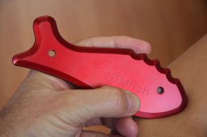
Care of your STMfish®
Your STMfish® is a precision instrument. Careless use can damage the highly polished working edge of the instrument.
- Clean with baby or alcohol wipes between usage.
- As with any IASTM tool, avoid bumping the edges against other metallic or hard surfaces as impacts can deform the working edge.
- When not in use, the instrument should be cleaned with alcohol and kept in the optional pouch or wrapped in a soft cloth.
Minor edge damage
In the event of careless use or accidental damage, small nicks and dents can be repaired with little effort or time. Inspect your STMfish® instrument from time to time by running your fingernail along the working edge.
Materials needed for repair are; silver polish, a soft cloth and "wet & dry" abrasive sheets of 400, 600, 800, 1000 and 1200 grit. Be aware that repairs to the Aluminium STMfish will remove anodising.
Take a wetted sheet of 400 grit waterpaper and work the damaged area of the tool until the working edge is smooth. Flush regularly with clean water and continue working the edge until the nicks cannot be detected with your fingernail. Then progress to 600, 800, 1000 and 1200 grit paper. When the surface is smooth finish off by polishing with your silver polish.
NB: Do not use strong caustic cleaning agents or products containing ammonia on the anodised aluminium STMfish as they will damage the anodising.
-
Basic STMfish® technique
Before using the instrument please read the cautions page.
Although the instrument can be used by applying the principles of manual soft tissue techniques as taught in professional manual therapy courses, training specific to the use of instrument assisted soft tissue mobilisation (IASTM) or Gua Sha is required.
Before using the STMfish, the area may be treated with hot packs and the skin washed thoroughly. The skin should then be carefully examined for any evidence of lesions or conditions that may be contra-indicative of IASTM therapy (see Cautions page).
A wide variety of lubricating media, ranging from organically based sorbolene creams to proprietary massage oils or waxes can also be used. Generally low viscosity oils and creams provide low traction and thicker creams and waxes create higher tissue traction. In excessively hairy body areas, a disposable square of fine cotton or satin fabric may be used as the "lubricating" layer between instrument and skin.
The most common error is the use of excessive pressure which can lead to significant bruising or tissue trauma. Be patient: Do not try to reduce chronic adhesions in 1 go!
The basic technique of using the STMfish® involves 5 important steps:
- Scanning the injured area to identify the extent and degree of injury.
- Assessing areas associated by fascial trains both proximal and distal to the focal injury. If you are not familiar with this anatomy, Thomas Myers book "Anatomy Trains: Myofascial Meridians for Manual and Movement Therapists" is an excellent resource.
- Determining the direction, depth and length of stroke required to release the affected tissues.
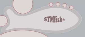
- Selecting the appropriate STMfish® profile to suit the area of treatment.
- Starting with the most superficial fascial layers slowly progress to deeper layers.
Scanning
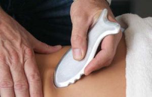
Use the instrument to apply your selected lubricating medium to minimise contact trauma. Scanning is mostly performed with a broad non focal contact employing very light long strokes over the area of concern. The weight of the stainless steel instrument is almost enough pressure. Select the STMfish® profile that provides the largest contact area. Feel for "gritty" vibrations that can indicate adhesions and scar tissue in the area examined.
Treatment Strokes
Once scanning has identified areas requiring release. Start with light medium (5-10cm) strokes, feeling for loss of subcutaneous "glide". Repeat strokes in the direction where adhesions are most evident until maximal float of the superficial tissue is restored.
Where marked adhesions occur, change to shorter (1-2cm) strokes with a smaller edge profile, slowly changing direction until you have gone through 360 degrees. Spend more time in the restricted directions and gradually work down through the tissue layers.
Modified Pin & Stretch technique is very useful for deeper structures. You can employ active or passive lengthening of the involved muscle or tendon. Put the muscle associated with the target tissue in a shortened position, and then place the instrument distal to the injured area to pin the tissue while slowly stretching the distal end of the muscle.
Lymphatic Drainage
During and after focal treatment strokes, apply light long strokes in the direction of natural lymphatic drainage of the injured area. The wave edge can be used where there is interstitial oedema present. This creates areas of high and low pressure "corrugations" in the tissue to stimulate the lymph vessels.
To sum up, The objectives of treatment are
- to release adhesions between fascial layers,
- to break down poor quality scar tissue in the underlying structures,
- to stimulate the repair of damaged tissue,
- to promote blood flow to areas of injury ,
- to enhance lymphatic drainage from injured tissue.
Remember IASTM is only part of the overall process of healing. Practitioners still need to attend to their primary professional management and active rehabilitation advice for the best results.
Recommended Reading
- Thomas Myers; Anatomy Trains: Myofascial Meridians for Manual and Movement Therapists
- Craig Liebenson; Rehabilitation of the Spine - a Practitioners Manual
- Kendall et al; Muscles - Testing and Function with Posture and Pain (Contains a useful DVD that reviews tissue layers)
- Hoppenfeld; Physical Examination of the Spine and Extremities
- Michael Schuenke; General Anatomy and the Musculoskeletal System (THIEME Atlas of Anatomy)
- Philipp Richter & Eric Hebgen; Trigger Points and Muscle Chains in Osteopathy